
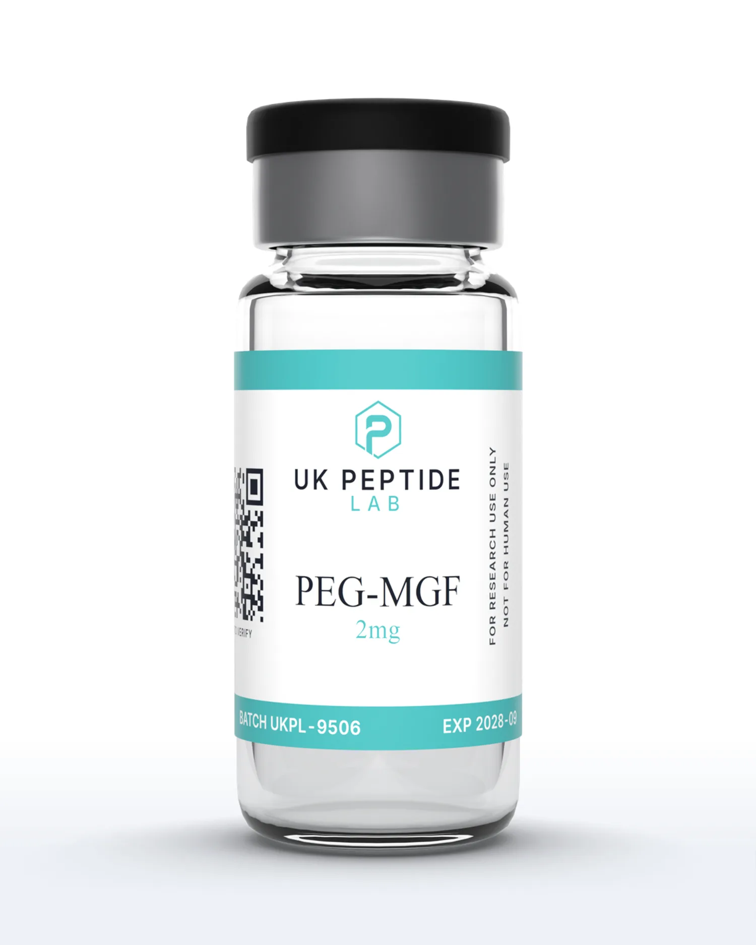
PEG-MGF
2mg
PEG-MGF is a PEGylated form of mechano growth factor, the IGF-1Ec splice-variant peptide expressed in muscle tissue in response to mechanical load. PEGylation of the 24-residue sequence slows clearance relative to the unmodified peptide, and research has examined both forms in models of muscle cell proliferation and satellite cell activation. Supplied as lyophilised powder, supplier-tested, with the certificate published on this page and independent third-party verification pending.
Published research on PEG-MGF is indexed on PubMed.
Intended Use: Strictly for in-vitro laboratory research. Not for use in humans or animals. Not to be used in foods, drugs, or medical diagnostics. This product has not been evaluated by the MHRA or FDA. Buyer assumes full responsibility for safe handling and regulatory compliance.
TBC
Purity
LAB
Certified
SAME
Day Ship
Quantity
Add £15.01 more for free UK delivery
Damaged or lost? Free reshipment guaranteed
Bacteriostatic Water 10ml
Required to reconstitute lyophilised peptides.
Research Use Only
This product is intended strictly for research and laboratory use only. Not for human consumption, medical use, or diagnostic purposes.
Certificate of Analysis
Third-party tested
Lyophilised Stability
24 months
Sealed at -20°C, light-protected
Reconstituted Stability
Up to 3 months
Stored at 2-8°C after mixing
Cold-Chain Shipping
Always
Dry-cold pack, Royal Mail Tracked 24
Important Notice
All compounds are sold individually and do not include research supplies. Products are provided in lyophilised (powder) form and require proper reconstitution before use in research settings.
Related Products
PEG-MGF Price (UK)
PEG-MGF 2mg is £29.99 per vial, supplied from stock in the UK with a third-party HPLC certificate published on this page. UK delivery is £4.50 by Royal Mail Tracked 24, or free once your basket reaches £45 (£15.01 away at this price). Orders placed before 2pm on a working day are dispatched the same day.
| Vial | Price | Cost per mg | Availability |
|---|---|---|---|
| PEG-MGF 2mgThis page | £29.99 | £14.99 | In stock |
Prices are in GBP and shown per vial as supplied, lyophilised, for in-vitro laboratory research only. Cost per mg is given so researchers can compare vial sizes on a like-for-like basis. Stock and pricing on this table are read live from our inventory, so they match the basket. See our FAQ for delivery and payment options, and quality & testing for how each batch is verified.
Product Specifications
Research Overview
PEG-MGF is the PEGylated form of mechano growth factor (MGF), the 24-residue C-terminal E-domain peptide of the IGF-1Ec splice variant of insulin-like growth factor 1. The underlying peptide sequence is Tyr-Gln-Pro-Pro-Ser-Thr-Asn-Lys-Asn-Thr-Lys-Ser-Gln-Arg-Arg-Lys-Gly-Ser-Thr-Phe-Glu-Glu-His-Lys, written YQPPSTNKNTKSQRRKGSTFEEHK, with a molecular weight of approximately 2866 g/mol unmodified and approximately 9.8 kDa in the PEGylated form supplied here. The splice variant was first cloned from mechanically stretched rabbit muscle by Yang, Alnaqeeb, Simpson and Goldspink (J Muscle Res Cell Motil, 1996), and its expression in muscle in response to mechanical load is what the mechano growth factor name records. PEG-MGF is a conjugate rather than a discrete small molecule and carries no CAS registry number on this listing.
The splice biology matters because it defines what the molecule is. The IGF-1Ec transcript carries a 49-base insert in the human sequence, and because the insert is not a multiple of three, the reading frame shifts and the C-terminal E domain is encoded in a different frame from the systemic IGF-1Ea isoform. It is that E-domain peptide, rather than the mature IGF-1 protein, that the early literature connected to muscle repair: the peptide drives myoblast proliferation while blocking differentiation (Yang and Goldspink, FEBS Lett, 2002), appears as an early pulse after local muscle damage in association with satellite cell activation (Hill and Goldspink, J Physiol, 2003), and increased progenitor cell numbers in cultures from normal, dystrophic and ALS muscle (Ates et al., FEBS Lett, 2007). Goldspink's review (Physiology, 2005) frames MGF as the local, damage-triggered isoform, with splicing switching toward IGF-1Ea as repair proceeds.
The PEGylation step is a pharmacokinetic modification, not a change to the peptide chemistry. The unmodified E peptide is cleared from circulation within minutes, so conjugation of polyethylene glycol, raising the mass to roughly 9.8 kDa, is designed to slow clearance through increased hydrodynamic radius, reduced renal filtration and shielding from proteases, the standard logic of the field reviewed by Harris and Chess (Nat Rev Drug Discov, 2003). In primary human muscle cell culture, the unmodified E peptide increased proliferation and delayed senescence in cultures from neonatal and young adult donors but not old adult muscle (Kandalla et al., Mech Ageing Dev, 2011), and in a mouse transplantation model it improved myogenic precursor cell engraftment (Mills et al., Am J Transplant, 2007).
It is worth being direct about the state of the evidence. The primary literature concerns the unmodified E-domain peptide, and the PEGylated form itself has no dedicated peer-reviewed publications, so its behaviour in any assay is an extrapolation from the unmodified molecule and from general PEGylation pharmacology. The founding in-vitro claims have also been challenged directly: a study conducted at two pharmaceutical companies found no effect of the MGF C-terminal peptide, at concentrations up to 500 ng/ml, on the proliferation or differentiation of C2C12 cells, primary human myoblasts or primary mouse muscle stem cells, and concluded that the physiological role of MGF itself should be questioned (Fornaro et al., Am J Physiol Endocrinol Metab, 2014). Human evidence is thin to absent, and the field's own review literature describes MGF as a putative repair factor (Matheny, Nindl and Adamo, Endocrinology, 2010). PEG-MGF is not an approved medicine in any jurisdiction and is supplied as lyophilised powder, supplier-tested at 99.77% purity with batch UKPL-9506, with the certificate published on this page and independent third-party verification pending, for in-vitro laboratory research only.
Product Specifications
Laboratory Handling
- Store lyophilised vials at -20°C, protected from light and moisture. Reconstituted PEG-MGF should be refrigerated at 2-8°C and used within 4 weeks.
- Reconstitute with bacteriostatic water, injecting the solvent slowly down the inner glass wall rather than onto the powder, then swirl gently until fully dissolved. Do not shake or vortex, and avoid vigorous agitation of reconstituted PEG conjugates.
- The 24-residue E-domain sequence contains no methionine, cysteine or tryptophan, so there are no oxidation-prone residues or disulfides to manage; the handling considerations are the PEG moiety (hydrophilic, altered behaviour on filters and gels) and sterility.
- Handle under aseptic laboratory conditions with alcohol-swabbed stoppers and sterile syringes. Avoid repeated freeze-thaw cycles and prepare measured aliquots from reconstituted stock for consistent experimental use.
Frequently Asked Questions
What is PEG-MGF?
PEG-MGF is the PEGylated form of mechano growth factor, the 24-residue E-domain peptide of the IGF-1Ec splice variant of insulin-like growth factor 1, sequence Tyr-Gln-Pro-Pro-Ser-Thr-Asn-Lys-Asn-Thr-Lys-Ser-Gln-Arg-Arg-Lys-Gly-Ser-Thr-Phe-Glu-Glu-His-Lys, written YQPPSTNKNTKSQRRKGSTFEEHK. The unmodified peptide has a molecular weight of approximately 2866 g/mol; the PEGylated conjugate supplied here runs at roughly 9.8 kDa, and as a conjugate rather than a discrete small molecule it carries no CAS registry number on this listing. The splice variant itself was cloned from rabbit muscle subjected to mechanical stretch by Yang, Alnaqeeb, Simpson and Goldspink (J Muscle Res Cell Motil, 1996), which is where the mechano growth factor name originates. One point of nomenclature matters to experimental design: MGF refers in the literature both to the full-length IGF-1Ec splice-variant protein and, in the peptide literature, to its C-terminal E-domain peptide alone. PEG-MGF is the latter, conjugated to polyethylene glycol. The PEGylation rationale is pharmacokinetic: the unmodified E peptide is cleared within minutes, and conjugation of polyethylene glycol increases hydrodynamic radius, reduces renal filtration and slows proteolytic degradation, the standard logic of the field reviewed by Harris and Chess (Nat Rev Drug Discov, 2003). Supplied as lyophilised powder for in-vitro laboratory research use only, not a licensed medicine anywhere.
PEG-MGF vs MGF
The two entries in this catalogue contain the same 24-residue E-domain peptide, YQPPSTNKNTKSQRRKGSTFEEHK; what separates them is the polyethylene glycol. The unmodified MGF E peptide is the molecule that appears throughout the primary literature, from the myoblast proliferation work of Yang and Goldspink (FEBS Lett, 2002), through the satellite cell activation studies of Hill and Goldspink (J Physiol, 2003), to the human primary muscle culture work of Kandalla and colleagues (Mech Ageing Dev, 2011). It is also the molecule whose clearance limits its utility, since the peptide disappears within minutes, which confines most published work to cell culture or local delivery. PEG-MGF carries the same sequence with polyethylene glycol conjugated to it, raising the mass from roughly 2866 g/mol to approximately 9.8 kDa. The trade is interpretability against residence time. The unmodified peptide maps directly onto the published literature, whereas the PEGylated form does not appear in the primary MGF literature at all, so its behaviour in any given assay is an extrapolation from the unmodified molecule and from general PEGylation pharmacology rather than an established finding. PEGylation also changes assay behaviour in ways that need controlling: the conjugate runs at higher apparent mass on size-exclusion chromatography, the polymer can interfere with antibody-based detection, and conjugation can alter receptor interaction. Researchers choose MGF when fidelity to the published molecule matters, and PEG-MGF when extended exposure is the design priority and the deviation from the literature is accepted as a variable.
PEG-MGF Research Applications
PEG-MGF inherits its research programme from the unmodified E-domain peptide, and every application below is drawn from the unmodified literature, because the PEGylated 24-mer itself has no dedicated peer-reviewed publications. The proliferation line: Yang and Goldspink (FEBS Lett, 2002) reported that the IGF-I Ec peptide stimulates proliferation of C2C12 myoblasts while blocking differentiation, the opposite balance to mature IGF-I, and Ates and colleagues (FEBS Lett, 2007) reported increased progenitor cell numbers in cultures derived from normal, dystrophic and ALS muscle. The satellite cell line: Hill and Goldspink (J Physiol, 2003) showed that the MGF splice variant is expressed as an early pulse after local muscle damage and is associated with satellite (stem) cell activation, and Kandalla and colleagues (Mech Ageing Dev, 2011) reported that the E peptide increased proliferation and delayed senescence in primary human muscle cell cultures from neonatal and young adult donors, though not from old adult muscle. The transplantation line: Mills and colleagues (Am J Transplant, 2007) reported that the E peptide improved myogenic precursor cell engraftment in a mouse model. The honest boundaries: a study conducted at two pharmaceutical companies found no effect of the MGF C-terminal peptide, at concentrations up to 500 ng/ml, on the proliferation or differentiation of C2C12 cells, primary human myoblasts or primary mouse muscle stem cells, and concluded that the physiological role of MGF itself should be questioned (Fornaro et al., Am J Physiol Endocrinol Metab, 2014); human evidence is thin to absent, with no clinical trials; and the field's own review literature describes MGF as a putative repair factor rather than an established one (Matheny, Nindl and Adamo, Endocrinology, 2010). Material supplied here is for in-vitro laboratory research only.
Reconstitution Guide
Allow the vial and the diluent to reach room temperature, then sterilise the rubber stopper of both the peptide vial and the bacteriostatic water vial with an alcohol swab. Slowly draw the bacteriostatic water into a sterile syringe and inject it gently down the inner glass wall of the vial, never directly onto the lyophilised powder. Swirl gently in a slow circular motion until fully dissolved; the solution should be clear and colourless. Never shake or vortex, and avoid vigorous agitation of reconstituted PEG conjugates. The underlying 24-residue sequence contains no methionine, cysteine or tryptophan, so the peptide itself carries no oxidation-prone residues and no disulfide chemistry; the handling consideration is the PEG moiety, which is hydrophilic and fully soluble but turns the conjugate into a polymer-sized species, so it behaves differently from a small peptide in membrane filtration and chromatographic steps. Work under sterile conditions, minimise stopper-open time, label the vial with the reconstitution date and refrigerate immediately.
See our full peptide reconstitution guide and reconstitution calculator for step-by-step protocol.
Storage Instructions
Store lyophilised vials at -20°C, protected from light and moisture, for long-term stability of up to 24 months. Once reconstituted with bacteriostatic water, hold the solution at 2-8°C (standard refrigerator) and use within approximately 4 weeks. For longer storage, prepare single-use aliquots at -20°C and thaw each aliquot only once; repeated freeze-thaw cycles are the fastest route to aggregation, and PEG conjugates are not exempt from this. Keep vials tightly sealed with minimal headspace, protect from light, and never store the solution in contact with oxidising agents or organic solvents. Handle under sterile laboratory conditions throughout, and keep the batch number and certificate of analysis on file for experimental records. For in-vitro laboratory research use only.
Frequently Asked Questions
Research References
- Different roles of the IGF-I Ec peptide (MGF) and mature IGF-I in myoblast proliferation and differentiation (Yang and Goldspink, FEBS Lett, 2002)
- Expression and splicing of the insulin-like growth factor gene in rodent muscle is associated with muscle satellite (stem) cell activation following local tissue damage (Hill and Goldspink, J Physiol, 2003)
- Mechano Growth Factor E peptide (MGF-E), derived from an isoform of IGF-1, activates human muscle progenitor cells and induces an increase in their fusion potential at different ages (Kandalla et al., Mech Ageing Dev, 2011)
- Mechano-growth factor peptide, the COOH terminus of unprocessed insulin-like growth factor 1, has no apparent effect on myoblasts or primary muscle stem cells (Fornaro et al., Am J Physiol Endocrinol Metab, 2014)
Related Research Compounds
PEG-MGF
2mg · £29.99